
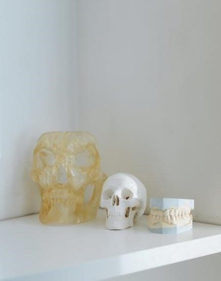
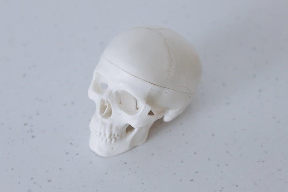
The skull is the skeleton of the head, comprising 22 bones that protect the brain and form cavities for sensory organs. It includes cranial and facial skeletons.
Overview of Skull Anatomy
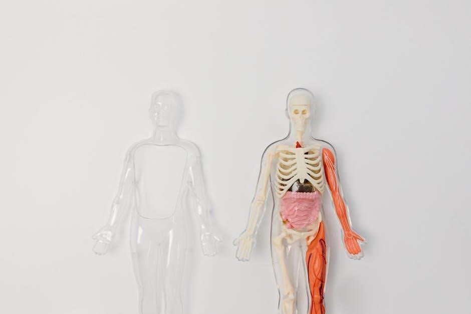
The skull, or cranial skeleton, is a complex structure comprising 22 bones that form the framework of the head. It is divided into two main sections: the cranial skeleton, which encases the brain, and the facial skeleton, which supports the face and its sensory organs. The cranial skeleton consists of 8 bones, including the frontal, parietal, occipital, temporal, sphenoid, and ethmoid bones, forming the cranial cavity. The facial skeleton includes 14 bones, such as the maxillary, zygomatic, and nasal bones, creating cavities for the eyes, nose, and mouth. The skull also features various foramina, openings that allow nerves and blood vessels to pass through. Understanding skull anatomy is essential for medical and anatomical studies, providing insights into human evolution, development, and clinical applications.
Importance of Studying Skull Anatomy
Studying skull anatomy is crucial for understanding the structural foundation of the head and its functional significance. The skull protects the brain, houses sensory organs, and provides attachment points for muscles and fasciae. Knowledge of skull anatomy aids in diagnosing and treating neurological and facial disorders, such as fractures or tumors. It also informs surgical approaches, including neurosurgery and reconstructive procedures. Additionally, skull anatomy is vital for understanding human evolution and variation. Forensic anthropology relies on skull features for identification purposes. For educators and students, detailed anatomical studies enhance learning and clinical preparedness. Overall, skull anatomy is a cornerstone of medical and biological sciences, offering insights into both health and history.
Brief History of Skull Anatomy Studies
The study of skull anatomy dates back to ancient civilizations, with early contributions from Egyptian and Greek physicians. Galen and Vesalius later detailed skull structures, laying the groundwork for modern anatomy. In the 19th century, anatomists like Gray and Huxley expanded knowledge, while radiographic imaging in the 20th century revolutionized skull analysis. Today, advanced techniques like CT scans and MRI provide detailed insights. Historical studies have also explored skull variations in evolution and forensic contexts. Understanding the historical progression of skull anatomy highlights its significance in medicine, anthropology, and neuroscience, offering a foundation for contemporary research and clinical applications.
Structure of the Skull
The skull is divided into two main sections: the cranial skeleton and the facial skeleton. It forms a protective framework for the brain and supports facial structures, creating cavities for sensory organs.

Cranial Skeleton

The cranial skeleton, or neurocranium, forms the protective framework of the skull, enclosing the brain. Comprising eight bones—frontal, parietal, occipital, temporal (2), sphenoid, and ethmoid—it creates the cranial cavity. These bones fuse during development, forming a rigid structure. The cranial base, part of the neurocranium, includes the foramen magnum, a critical opening for the spinal cord. Its intricate design ensures brain protection while accommodating cranial nerves and blood vessels. The sphenoid and ethmoid bones contribute to the cranial base’s complexity, forming part of the orbital and nasal cavities. This skeleton’s structure is vital for both protection and functional support of the central nervous system, making it a cornerstone of skull anatomy studies and clinical applications.
Facial Skeleton
The facial skeleton consists of 14 bones forming the framework of the face, orbits, and nasal cavities. It includes the maxillary, zygomatic, nasal, lacrimal, palatine, and inferior nasal conchae bones. These bones create cavities for the eyes, nose, and mouth, while also anchoring muscles for facial expression. The maxillary bones, the largest, form the upper jaw and part of the orbital floor. The zygomatic bones shape the cheekbones and contribute to the lateral orbital walls. The nasal bones form the bridge of the nose, while the lacrimal bones house tear ducts. Palatine bones form the hard palate, separating nasal and oral cavities. Together, these bones provide structural support and facilitate sensory functions, making the facial skeleton vital for both aesthetics and physiology. Its detailed anatomy is crucial in surgical and reconstructive procedures, as highlighted in anatomical studies and clinical notes.
Craniofacial Junction
The craniofacial junction is the transitional zone between the cranial and facial skeletons, playing a pivotal role in skull anatomy. It involves the connection of the frontal bone, sphenoid bone, and temporal bones, forming a complex structural and functional interface. This junction supports the brain’s frontal lobe and temporal lobe, while also housing critical neurovascular structures. The sphenoid bone, wedged between the frontal and temporal bones, is central to this junction, providing anchorage for dural folds like the tentorium cerebelli. The foramen magnum, located nearby, allows passage of the spinal cord. This area is vital for neurosurgical approaches and understanding craniofacial development. Its intricate anatomy is emphasized in clinical notes and studies, highlighting its significance in both anatomical and surgical contexts. Proper comprehension of this junction is essential for diagnosing and treating craniofacial abnormalities and trauma.
Bones of the Skull
The skull comprises 22 bones, divided into cranial and facial bones, forming a protective and functional framework for the brain and sensory organs, with unique morphometric variations.
Cranial Bones
The cranial bones form the cranial skeleton, which creates the cranial cavity housing the brain. There are 8 cranial bones: frontal, parietal (2), occipital, temporal (2), sphenoid, and ethmoid. These bones are fused in adults, forming a rigid structure. The frontal bone forms the forehead and part of the eye sockets. The parietal bones make up the roof and sides of the skull. The occipital bone is at the back, containing the foramen magnum, which connects to the spinal cord. The temporal bones house structures for hearing and balance. The sphenoid and ethmoid bones are complex, with air-filled cavities. These bones ossify from both intramembranous and endochondral ossification. Their morphology varies, with clinical significance in surgery and neurological studies.
Facial Bones
The facial bones constitute the facial skeleton, comprising 14 bones that form the structure of the face. These bones include the maxillary, zygomatic, nasal, lacrimal, palatine, and inferior nasal conchae bones. The maxillary bones form the upper jaw and palate, while the zygomatic bones shape the cheeks. The nasal bones create the bridge of the nose, and the lacrimals house tear ducts. The palatine bones contribute to the hard palate and nasal cavity, and the inferior nasal conchae are spongy bones within the nasal passages.
These bones vary in shape and function, supporting facial features and forming cavities for sensory organs. Their morphology is crucial in clinical contexts, such as reconstructive surgery and trauma management.
Classification of Skull Bones
The skull bones are classified into cranial bones and facial bones. Cranial bones form the cranial skeleton, protecting the brain, while facial bones shape the face. Cranial bones include frontal, parietal, occipital, temporal, sphenoid, and ethmoid bones. Facial bones consist of maxillary, zygomatic, nasal, lacrimal, palatine, and inferior nasal conchae bones. These bones are further categorized as paired (e.g., parietal, temporal) or unpaired (e.g., frontal, occipital). They also differ in their fusion; cranial bones fuse into a single unit, while facial bones remain more articulated. This classification aids in understanding their roles in protection, sensory function, and structural support. Their unique morphologies and positions are critical in anatomical and clinical studies.

Cranial Bones
The cranial bones form the cranial skeleton, protecting the brain. They include frontal, parietal, occipital, temporal, sphenoid, and ethmoid bones, fused to create a sturdy structure.
Frontal Bone
The frontal bone forms the forehead and upper eye sockets, protecting the brain’s frontal lobe. It is a single bone with a squama frontalis (upper part) and a pars orbitalis (orbital section). The frontal bone contains air-filled cavities known as frontal sinuses, which vary in size and shape among individuals. It articulates with the nasal bones and maxillary bones at the nose and with the parietal bones at the vertex of the skull. The frontal bone also features a supraorbital ridge, providing attachment points for muscles and forming the brow. Its structure is crucial for both the cranial and facial skeletons, contributing to the skull’s overall integrity and aesthetic features.
Parietal Bones
The parietal bones are a pair of flat, curved bones located on the sides and roof of the skull. They form part of the cranial vault, which protects the brain. Each parietal bone has a squamous part (flat portion) and a vertical part (thicker, more robust section). The bones articulate with the frontal bone anteriorly, the occipital bone posteriorly, and the temporal bones inferiorly. The parietal tuberosity is a raised area on the outer surface for muscle attachment. Internally, the bones feature the superior and inferior parietal gyri, which correspond to brain convolutions. The parietal bones fuse during adulthood, forming a single structure. Their thickness varies, providing structural support and protection to the cranial cavity.
Occipital Bone
The occipital bone is located at the posterior and inferior part of the skull, forming the back and base of the cranium. It is a flat, trapezoidal bone with a large, oval opening called the foramen magnum, through which the spinal cord connects to the brain. The occipital bone articulates with the parietal bones superiorly and the temporal bones laterally, completing the cranial vault. It also forms part of the cranial base, supporting the brain. The bone features the external occipital crest, a ridge for muscle attachment, and the internal occipital crest, which houses the transverse sinuses. The occipital bone is essential for protecting the brain and providing structural support to the skull’s posterior region.
Temporal Bones
The temporal bones are a pair of irregular bones located on the lateral sides of the skull, playing a crucial role in both the cranial and facial skeletons. Each temporal bone forms part of the cranial base and houses the temporomandibular joint, essential for jaw movement. The bone features the external acoustic meatus, which connects to the ear canal, and the mastoid process, a bony projection behind the ear. The styloid process extends downward from the temporal bone, serving as an attachment point for muscles and ligaments. The temporal bone also contains several foramina, including the foramen spinosum (transmitting the middle meningeal artery) and the stylomastoid foramen (allowing the facial nerve to exit the skull). Its complex structure supports auditory function, cranial protection, and facial anatomy.
Sphenoid Bone
The sphenoid bone is a butterfly-shaped bone located in the cranial base, connecting the cranial and facial skeletons. It supports the frontal lobe and parts of the temporal lobes, forming a bridge between these regions. The bone consists of a central body and two pairs of wings (greater and lesser), which extend outward and upward. The body of the sphenoid forms part of the cranial cavity and contains the sphenoidal sinuses, air-filled cavities that reduce skull weight. Key features include the foramen ovale and foramen spinosum, which transmit nerves and blood vessels. The sphenoid bone plays a critical role in cranial structure, supporting the brain and facilitating nerve passage, making it essential for both anatomical integrity and functional processes.
Ethmoid Bone
The ethmoid bone is a spongy, irregular bone located between the nasal cavity and the eye sockets; It forms part of the anterior cranial base and the roof of the nasal cavity. The bone is characterized by its lightweight, spongy structure due to numerous air cells. Key features include the cribriform plate, which is perforated by olfactory nerves, and the perpendicular plate, forming part of the nasal septum. The ethmoid bone also contributes to the medial wall of the orbit and connects with the frontal, sphenoid, and lacrimal bones. Its air cells are part of the paranasal sinus system, aiding in reducing skull weight while maintaining structural integrity. This bone is vital for separating cranial and facial cavities while supporting sensory functions.
Facial Bones
The facial bones form the structure of the face, protecting sensory organs and creating cavities for the eyes, nose, and mouth. They include the maxillary, zygomatic, nasal, and mandible.
Maxillary Bones
The maxillary bones are a pair of large, irregular bones forming the upper jaw and part of the facial skeleton. They are crucial for supporting the face and forming cavities for the eyes, nose, and mouth. Each maxillary bone contains the maxillary sinus, the largest paranasal sinus, which varies in size and shape among individuals. These bones articulate with the frontal, zygomatic, and palatine bones, contributing to the structure of the orbital and nasal cavities. The maxillary bones also anchor the upper teeth and play a key role in facial aesthetics and function. Their complex anatomy makes them significant in both anatomical studies and clinical practices, particularly in surgeries involving the face and sinuses.
Zygomatic Bones
The zygomatic bones, also known as cheekbones, are a pair of irregular bones forming part of the lateral wall of the orbit and the floor of the orbit. They contribute to the structure of the face and play a key role in forming the prominence of the cheeks. Each zygomatic bone articulates with the frontal, maxillary, sphenoid, and temporal bones, forming part of the orbital and facial framework. The zygomatic arch, formed by the zygomatic and temporal bones, is a significant landmark for muscle attachment, particularly for muscles of mastication. The zygomatic bones also house the zygomaticofacial and zygomaticotemporal canals, which transmit nerves and blood vessels. Their complex anatomy makes them crucial in facial reconstruction surgeries and trauma management, as they are often involved in facial injuries due to their exposed position.
Nasal Bones
The nasal bones are two small, rectangular bones located at the bridge of the nose, forming the upper part of the nasal cavity. They articulate with the maxillary bones anteriorly and the frontal bone superiorly, while their posterior ends connect with the ethmoid bone. These bones contribute to the formation of the anterior cranial fossa and the roof and lateral walls of the nasal cavity. The nasal bones are vital for supporting the nasal structure and protecting the underlying olfactory organs; They also serve as attachment points for cartilaginous nasal septum and muscles of the nose. Their position and structure make them prone to fractures in facial trauma. The nasal bones are supplied by branches of the ophthalmic and maxillary arteries and are innervated by the nasociliary nerve, highlighting their importance in both sensory and structural functions of the face.
Lacrimal Bones
The lacrimal bones are the smallest and thinnest bones in the human skull, forming part of the facial skeleton. They are located in the medial wall of the orbit and contribute to the anterior portion of the nasal cavity. Each lacrimal bone has a distinctive groove, the lacrimal sulcus, which, together with the maxillary bone, forms the lacrimal fossa. This fossa houses the lacrimal sac, a structure critical for tear drainage. The lacrimal bone also features the lacrimal hamulus, a small hook-like projection that helps anchor the lacrimal sac. These bones articulate with the maxillary, palatine, and ethmoid bones, playing a key role in the structural integrity of the orbit and nasal cavity. Their involvement in the nasolacrimal duct system makes them essential for tear drainage and ocular health. Fractures or malformations of the lacrimal bones can lead to tear duct obstruction and other ophthalmological issues.
Palatine Bones
The palatine bones are paired bones located in the facial skeleton, forming part of the nasal cavity, orbit, and hard palate. They are situated posterior to the maxillary bones and contribute to the lateral walls of the nasal cavity. Each palatine bone consists of a horizontal plate and a vertical plate. The horizontal plate forms part of the hard palate, while the vertical plate contributes to the orbital and nasal walls. The palatine bones articulate with the maxillary, sphenoid, and ethmoid bones, as well as the inferior nasal conchae. They contain the palatine foramina, which transmit nerves and blood vessels. These bones are crucial for forming the posterior nasal apertures and supporting the structures of the face and skull. Their intricate articulations make them essential for maintaining craniofacial integrity and function.
Inferior Nasal Conchae
The inferior nasal conchae are long, thin, curved bones located within the nasal cavity. They are separate from the superior and middle nasal conchae, which are part of the ethmoid bone. These bones project into the nasal passages, increasing the surface area for air filtration and humidification. Each concha is covered by mucous membrane and plays a key role in respiratory function. The inferior nasal conchae also help to direct airflow through the nasal passages. They articulate with the maxillary bones, palatine bones, and lacrimal bones, forming part of the lateral nasal wall. Their structure is crucial for olfactory function and overall respiratory health, making them an essential component of the facial skeleton.
Landmarks and Features of the Skull
The skull features the Frankfurt plane, an anatomical reference, and the foramen magnum, a large opening at the base. Other notable landmarks include external and internal skull features.
External Landmarks
External landmarks of the skull include the Frankfurt plane, an anatomical reference line, and the external acoustic meatus, the opening of the ear canal. The mental protuberance is a ridge on the chin, while the zygomatic arches form the cheekbones. The occipital protuberance is a prominent bump at the back of the skull. These features are essential for identifying key anatomical reference points. They are visible on the surface and aid in locating other structural components. External landmarks are crucial for clinical examinations and surgical procedures, providing a framework for understanding skull anatomy. These features are easily palpable and serve as guides for various medical and anatomical studies. Their precise locations and shapes vary slightly among individuals but generally follow a consistent anatomical pattern.
Internal Landmarks
Internal landmarks of the skull include the foramen magnum, a large opening at the base through which the spinal cord connects to the brain. The tentorium cerebelli is a dural fold separating the cerebrum and cerebellum. The sella turcica houses the pituitary gland, while the cribriform plate contains olfactory nerve openings. These features are critical for neuroanatomy and surgery. Internal landmarks guide procedures like cranial surgeries and endoscopic interventions. They are not visible externally but are vital for understanding the skull’s internal structure. Their precise locations and relations to brain structures make them essential for medical professionals. These landmarks are studied in detail to ensure accurate diagnoses and treatments in neurological and surgical contexts.
Foramina of the Skull
The foramen magnum is the largest opening, transmitting the spinal cord and major blood vessels. Other significant foramina allow passage of cranial nerves and vascular structures, essential for neuroanatomy and surgery.
Foramen Magnum
The foramen magnum is the largest and most significant opening in the skull, located in the occipital bone. It serves as the passage for the spinal cord to connect with the brain, allowing vital nerve fibers and blood vessels to pass through. This foramen is crucial for maintaining neurological function and is a key anatomical landmark in neurosurgery and medical imaging. Its structure and position are essential for understanding cranial anatomy, particularly in procedures involving the brainstem and spinal cord connection. Damage to this area can lead to severe neurological deficits, emphasizing its importance in clinical studies and surgical approaches.
Other Significant Foramina
Beyond the foramen magnum, several other foramina play critical roles in skull anatomy. The superior orbital fissure transmits nerves and vessels to the eye, while the foramen rotundum allows the maxillary nerve to pass. The foramen ovale and foramen spinosum facilitate the passage of the mandibular nerve and middle meningeal artery, respectively. Additionally, the stylomastoid foramen enables the facial nerve to exit the skull. These foramina are essential for connecting cranial structures with external tissues, ensuring proper sensory and motor functions. Their precise locations and sizes are vital in neurosurgery and radiology, aiding in diagnostic and therapeutic interventions. Understanding these foramina is crucial for mapping cranial nerve pathways and avoiding complications during surgical procedures.

Clinical Significance of Skull Anatomy
Understanding skull anatomy aids in surgical planning, diagnostic accuracy, and preventing complications in neurosurgery and craniomaxillofacial procedures, ensuring precise interventions and improved patient outcomes.
Surgical Approaches
Surgical Approaches
Surgical approaches to the skull require precise knowledge of its anatomy to ensure safe and effective interventions. Understanding the cranial base, sphenoid bone, and temporal bones is critical for accessing neurological structures. Foramen Magnum serves as a key landmark for posterior fossa surgeries. Neurosurgical interventions, such as craniotomies, rely on identifying skull landmarks to minimize risks. Advanced imaging techniques, like CT scans, aid in preoperative planning. Mastery of skull anatomy enables surgeons to navigate complex regions, ensuring optimal outcomes. This knowledge is vital for treating conditions like tumors, aneurysms, and traumatic injuries, highlighting the importance of anatomical precision in surgical practice.
Neurological Implications
The skull’s anatomy has significant neurological implications, as it encases and protects the brain. The foramen magnum allows the spinal cord to connect with the brain, making it a critical landmark. Damage to skull bones, such as fractures, can compress or damage underlying neural structures. The middle meningeal artery, which runs through the temporal bone, plays a key role in supplying blood to the brain. Skull anomalies or malformations can lead to neurological deficits or increased risk of brain injuries. Understanding the skull’s relationship with the brain is essential for diagnosing and treating conditions like cerebral swelling, tumors, or traumatic injuries. Precise knowledge of skull anatomy aids in neurosurgical planning and ensures better patient outcomes.